
It was the summer of tenth grade when I was first introduced to fluorescent microscopy, and it was in the middle of my sophomore year when I started developing my interest in molecular/ systems biology in particular. I discovered early on how much the figures in a paper made up its data analysis and visualization: even if not all the words made sense, I had a sense of their essence when they were linked to a figure with images. I didn’t need all the details to understand what the paper was trying to say, but the fact that I understood it at all motivated me to continue fleshing out my knowledge of those details. They were similar to what I had learned in school and textbooks, but presented real experimental results that honed in on specific parts of cells.
I've always been a visual person–I sketch, paint, and do photography–and perhaps that was why I was so drawn to those images in particular. The forms and colors were arranged in an abstract and beautiful way. I remember enthusiastically showing a set of pictures in an article to a teacher at art camp during the winter of 10th grade. She wasn’t into biology at all, but she was curious about what the images were of after seeing them, we spoke about fluorescent proteins and what she remembered from high school biology.
My art teacher mentioned that if she had been introduced to the imaging aspect of biology sooner, she might have been more interested in it when she was younger, and that got me thinking. A lot of kids (myself included) have never used a microscope until high school, but I they’re a necessary tool for visualizing and therefore understanding biology. Even then, none of it was experimental: 9th grade biology was mainly observing pre-fixed samples on a glass microscope slide.
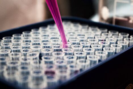
Now, I’m working on a project in Dr. Rafii’s lab at Weill Cornell exploring the role of PIEZO1, a mechanosensitive ion channel receptor, in vasculogenesis. I spend most of my time in the imaging room–a lot of my research involves taking z-stacked photos of wells in a plate or lanes in a microfluidic device throughout the development of a blood vessel. I work on selecting representative snapshots of a dataset, deconvolving confocal microscopy images, and using those to construct a 3D model of my sample to make my results as clear as possible. I also work with other members in the lab with their images of vessels vascularizing organoids or perfused by drugs or white blood cells, for example. Imaging is a major part of our work.
With all this in mind, my plan is to make presentations/ summaries of various pieces of literature in molecular biology with a focus on confocal microscopy images. My aim is to simplify the goals and results of these papers as much as possible in a way that’s both engaging and understandable for an elementary or middle schooler, using confocal imaging as a channel to help them visualize experimental biology. I want to provide a source of inspiration and introduce them to scientific biology in the hopes that a few of them, like me, would be interested in digging deeper.
More on Alison's outreach project to come!
I've always been a visual person–I sketch, paint, and do photography–and perhaps that was why I was so drawn to those images in particular. The forms and colors were arranged in an abstract and beautiful way. I remember enthusiastically showing a set of pictures in an article to a teacher at art camp during the winter of 10th grade. She wasn’t into biology at all, but she was curious about what the images were of after seeing them, we spoke about fluorescent proteins and what she remembered from high school biology.
My art teacher mentioned that if she had been introduced to the imaging aspect of biology sooner, she might have been more interested in it when she was younger, and that got me thinking. A lot of kids (myself included) have never used a microscope until high school, but I they’re a necessary tool for visualizing and therefore understanding biology. Even then, none of it was experimental: 9th grade biology was mainly observing pre-fixed samples on a glass microscope slide.
Now, I’m working on a project in Dr. Rafii’s lab at Weill Cornell exploring the role of PIEZO1, a mechanosensitive ion channel receptor, in vasculogenesis. I spend most of my time in the imaging room–a lot of my research involves taking z-stacked photos of wells in a plate or lanes in a microfluidic device throughout the development of a blood vessel. I work on selecting representative snapshots of a dataset, deconvolving confocal microscopy images, and using those to construct a 3D model of my sample to make my results as clear as possible. I also work with other members in the lab with their images of vessels vascularizing organoids or perfused by drugs or white blood cells, for example. Imaging is a major part of our work.
With all this in mind, my plan is to make presentations/ summaries of various pieces of literature in molecular biology with a focus on confocal microscopy images. My aim is to simplify the goals and results of these papers as much as possible in a way that’s both engaging and understandable for an elementary or middle schooler, using confocal imaging as a channel to help them visualize experimental biology. I want to provide a source of inspiration and introduce them to scientific biology in the hopes that a few of them, like me, would be interested in digging deeper.
More on Alison's outreach project to come!
